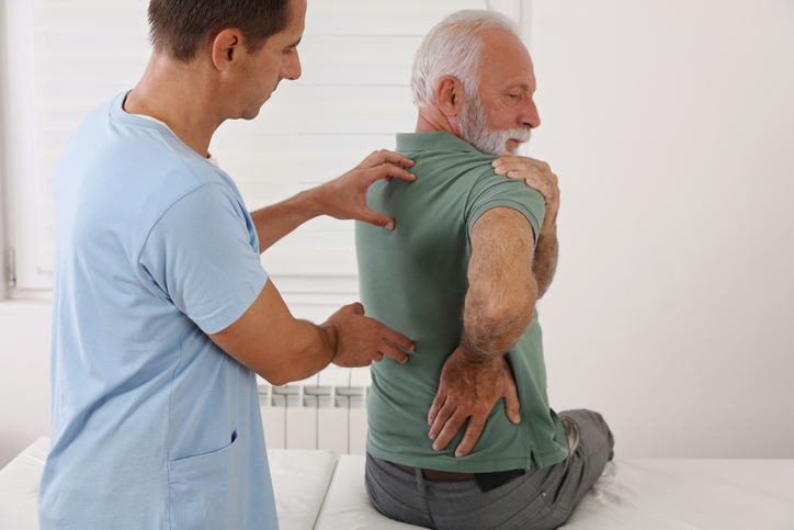
Pain
Anatomy of Spinal Stenosis

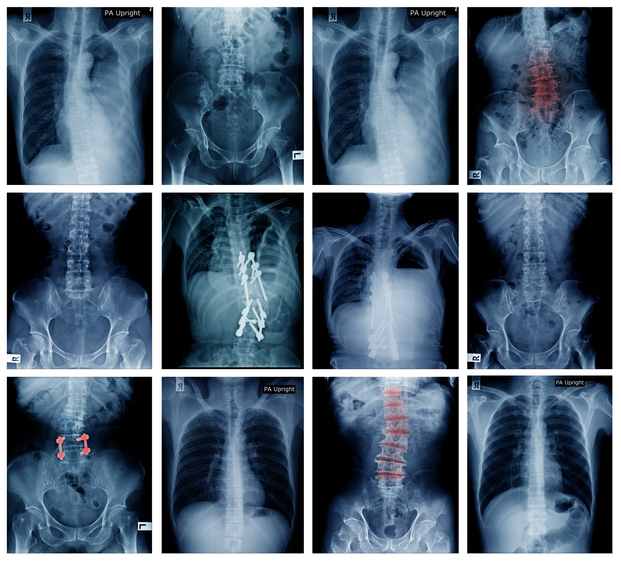
What is spinal stenosis?
Spinal stenosis is a narrowing of the spinal canal. It occurs when the bony openings within the spine (foramina) begin to narrow, placing pressure on the nerves traveling throughout the spine. This reduced nerve space can occur within the spinal cord or where the spinal nerves exit the spinal canal. Knowing the anatomy of the spine is beneficial in understanding spinal stenosis.
The spine
The spine, or backbone, runs from the neck to the lower back, close to the buttocks. The bones of the spine (vertebrae) form the spinal canal, which houses the spinal cord and nerves. The spine provides flexibility that allows a person to walk, twist, stand, bend and sit.
Vertebrae
Although a person is normally born with 33 vertebrae, certain ones at the bottom of the spine fuse together during growth. Therefore, the spine of an average adult consists of 24 vertebrae (bones). They connect with facet joints.
Facet joints
Facet joints are small joints located between each vertebra of the spine. They have cartilage to allow vertebrae to slide against each other. A pair of facet joints are located at the back of each spinal segment. These joints help support the spine and allow for range of motion.
Discs
Spinal discs are rubbery cushions that sit between individual vertebrae. They support the spine and keep the back pliable. These discs act as shock absorbers and have a soft, gel-like center. The outer ring is flexible. Due to constant pressure, a disc can tear, bulge or rupture.
Spinal cord and nerves
The spinal column consists of stacked bones (vertebrae). The spinal column extends from the skull to the lower back. The vertebrae house the spinal cord. The spinal cord is a column of nerves that run through the spinal canal. These nerves carry communication signals between the brain and the rest of the body. The spinal cord is responsible for many things including, but not limited to, transmitting pain and other sensory signals, allowing for movement (both voluntary and involuntary), and communicating with organs and organ systems.
Soft tissue
Soft tissues provide strength and stability that help with movements, such as twisting or bending. Soft tissue includes ligaments, muscles and tendons. Ligaments help connect bones, provide stabilization, and restrict movement outside a normal range. Muscles support the back and provide movement. Tendons connect bones and muscles together and help with movement.
Spine segments
The spine has five segments that begin at the neck and end close to the buttocks. These segments consist of the following:
- Neck or cervical — The top of the spine has seven vertebrae (C1 - C7) and makes an inward curve. It supports the weight of the head and is responsible for the ability to turn, tilt and nod the head.
- Middle back or thoracic — The chest area or thoracic spine consists of 12 vertebrae (T1 - T12) and bends slightly backwards. This section attaches to the ribs.
- Lower back or lumbar — The lower back is made up of five vertebrae (L1 - L5) and bears most of the body’s weight. The lumbar segment of the spine is typically responsible for lifting or carrying items. The lower back bends slightly inwards. It supports the upper spine and connects to the pelvis. Many back injuries occur in this segment.
- Sacrum — The sacrum has five vertebrae (S1 - S5) and connects to the hips. This segment has no movement since fusing occurs prior to birth.
- Tailbone or coccyx — This segment has four vertebrae and is located at the bottom of the spine. These vertebrae also fuse together and attach to ligaments and muscles of the pelvic floor.