Pain
Diagnosing Lumbar Spinal Stenosis

What is spinal stenosis?
Spinal stenosis is a narrowing of the spinal canal. It occurs when the bony openings within the spine (foramina) begin to narrow, placing pressure on the nerves traveling throughout the spine. This reduced nerve space can occur within the spinal cord or where the spinal nerves exit the spinal canal.
The spine (backbone) runs from the neck to the lower back. The bones of the spine (vertebrae) form the spinal canal, which houses the spinal cord and nerves. If a spinal nerve or the spinal cord becomes compressed, pain, numbness, muscle weakness, or tingling may occur. Spinal stenosis most often occurs in the lower back (lumbar stenosis) or neck (cervical stenosis). Spinal stenosis of the upper/middle back (thoracic stenosis) is rare.
Diagnosing lumbar spinal stenosis
Osteoarthritis is the most common cause of lumbar spinal stenosis. Pain is frequently experienced in the back and legs. Additionally, numbness, tingling, cramping and weakness may occur in the legs. Diagnosing lumbar spinal stenosis involves a medical history, physical examination, and imaging tests.
Medical history
A health care professional will gather a health history that includes other conditions and current medications. Personal and family medical history will also be reviewed. Symptoms, including pain location and type of pain, should be detailed.
Physical examination
A complete physical exam will include the health care professional pressing on areas of the spine to determine if it is painful. The individual will be asked to bend in certain directions to check for pain or other symptoms. Balance will be inspected while walking. Leg and arm strength will also be evaluated.
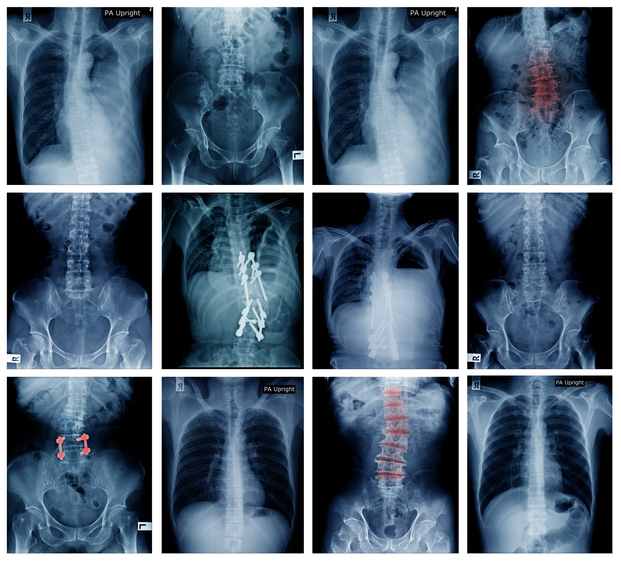
X-ray
An X-ray of the lumbar spine will show bone spurs and changes in bone structure. These can place pressure on spinal nerves, which causes narrowing of the spinal canal.
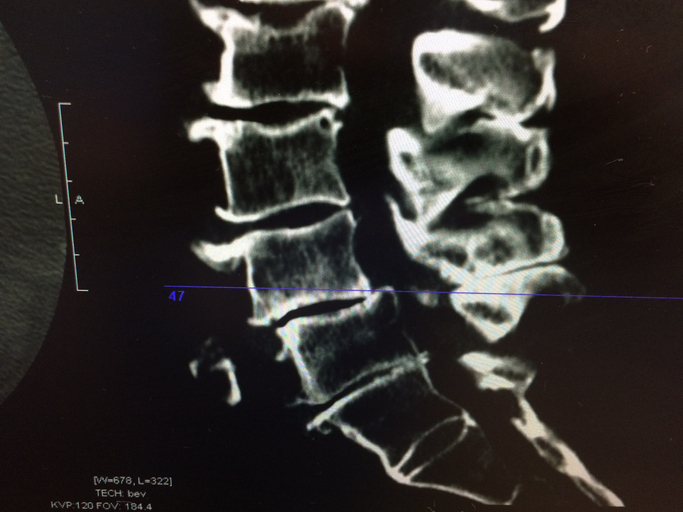
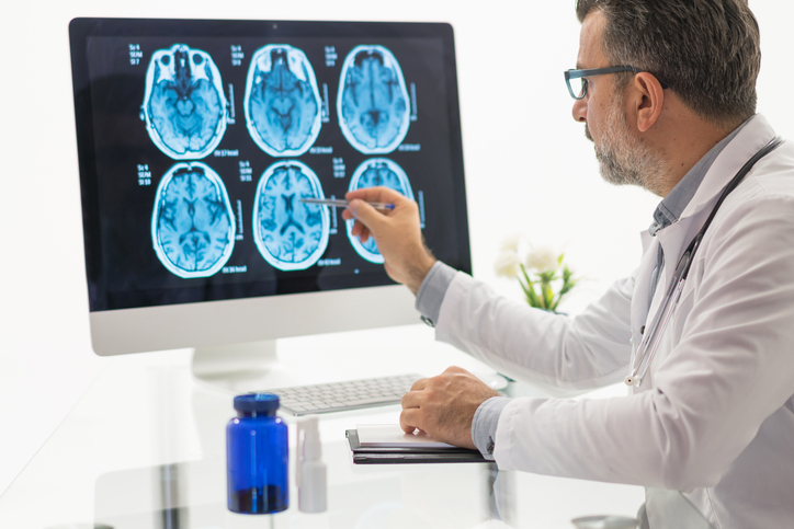
Imaging tests
A computed tomography (CT) scan uses X-rays to get a cross-sectional view of the spine. A CT myelogram involves the addition of a contrast dye to outline the spinal cord and nerves. A magnetic resonance imaging (MRI) shows detailed images by using a powerful magnet and radio waves. It can show tumors and damage to ligaments or discs.