Pain
Diagnosing Cauda Equina Syndrome
What is cauda equina syndrome?
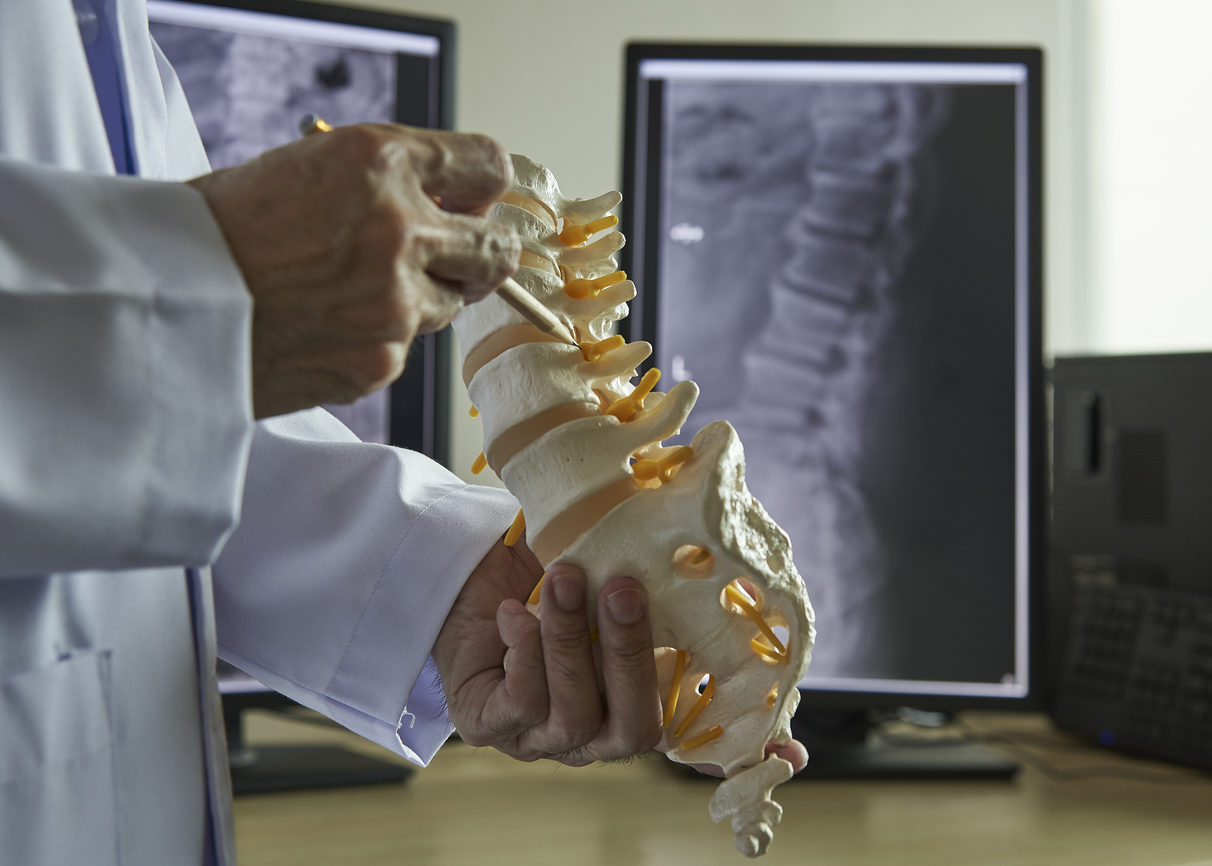
The cauda equina is a group of approximately 10 nerves and nerve roots located at the bottom of the spinal cord. They communicate with the brain to provide motor and sensory stimulation to the legs, pelvis, bladder, anus and bowels. Cauda equina syndrome (CES) is a rare but serious disorder that occurs when this nerve bundle becomes compressed or inflamed. CES typically results in a medical emergency that requires urgent surgical intervention to prevent the possibility of long-term paralysis, damage to organs in the pelvic region, neurological issues, difficulty walking, and other physical problems.
Diagnosing cauda equina syndrome
CES is oftentimes difficult to diagnose. However, if bowel, bladder and/or sexual difficulties are present, or paresthesia exists in the backs of the legs, buttocks, hip, and inner thighs, CES is typically diagnosed. A health care professional will review the medical history, perform a physical examination, and evaluate symptoms. Certain imaging tests may also be required.
Medical history
A medical history will be gathered and evaluated. This should include detailed symptoms, when symptoms began, symptom duration, description of pain, and pain severity. It will also include a family medical history, any recent falls, trauma or injuries, intravenous drug use, history of malignancy, chiropractic manipulation, and use of anticoagulant drugs.
Physical examination
A health care professional will check the reflexes, alignment, sensation, stability and strength. A review will be completed for standing, sitting, walking on the heels and toes, bending, and lying on the back while lifting the legs. Blood tests may also be ordered.
Imaging tests
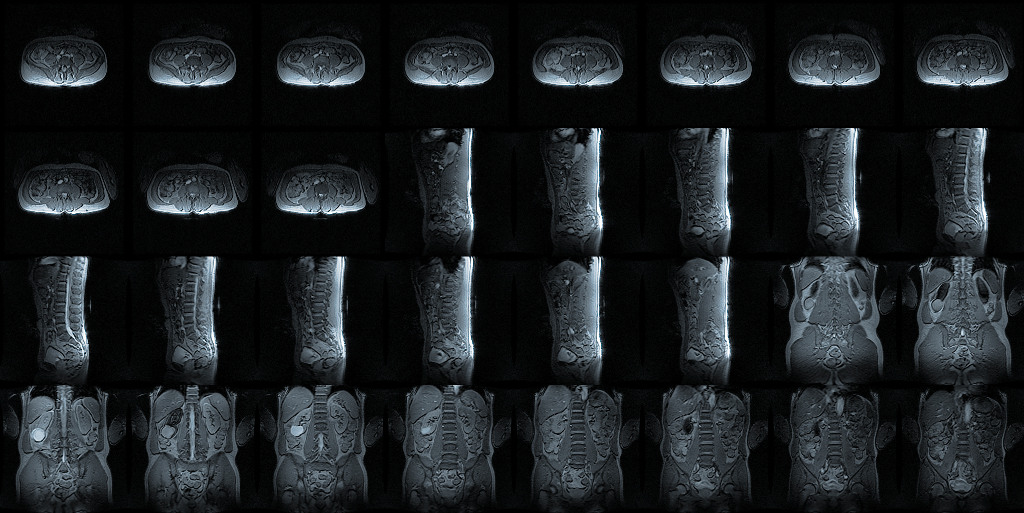
Other testing may be conducted to observe bones, spinal cord, nerve roots, herniated discs, bone spurs, or tumors. These testings include, but are not limited to, the following:
- A magnetic resonance imaging (MRI) scan uses magnetic fields to produce images of tissue around the spine and nerve roots.
- A myelogram imaging test involves a dye being injected into tissue around the spine to identify any spinal canal issues.
- An X-ray can show spinal cord issues or nerve compression from a herniated disk, tumor, or other problems.
- A computed tomography (CT) scan can help determine if an injury or disease is present. During a CT scan, X-rays and a computer are used to obtain a 3D image of bones and tissue.