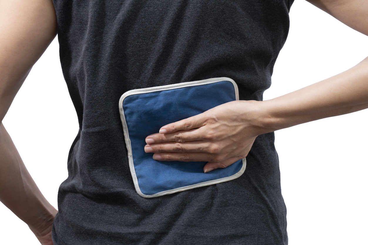
Pain
Diagnosing Spondylolysis

What is spondylolysis?
The term spondylolysis comes from a combination of two Greek words: spondylos (vertebra) and lysis (defect). Spondylolysis is a medical condition that involves a defect of the pars interarticularis. The pars interarticularis is a thin bone that hinges two vertebrae together in the lower back. Spondylolysis most commonly occurs in the lumbar vertebrae; however, it can occur in any section of the spine: cervical (neck), thoracic (mid back), or lumbar (lower back). Both cervical and thoracic spondylolysis are rare. If spondylolysis occurs in the cervical or thoracic spine, the equivalent structure of the pars interarticularis in that area of the spine is affected.
Diagnostic process
Spondylolysis most commonly develops in adolescents and is often asymptomatic. Diagnosing spondylolysis typically consists of a medical history review, physical examination, and imaging tests.
- Medical history
A health care professional will gather a medical history, review symptoms, and ask questions. Pain specifics will be discussed, including when the pain started, where it hurts, and if anything helps or makes it worse. - Physical examination
During a physical examination, a physician will perform the “one-legged hyperextension maneuver.” This involves standing on one leg in a position to extend the lumbar spine. This is repeated on the opposite leg. If pain occurs, it can be indicative of spondylolysis. A physician will analyze range of motion, muscle weakness, muscle spasms, and pain or tenderness in areas typically affected by spondylolysis (back, glutes, and thighs). They will also examine the gait for stiff walking and posture for awkward standing. - Imaging
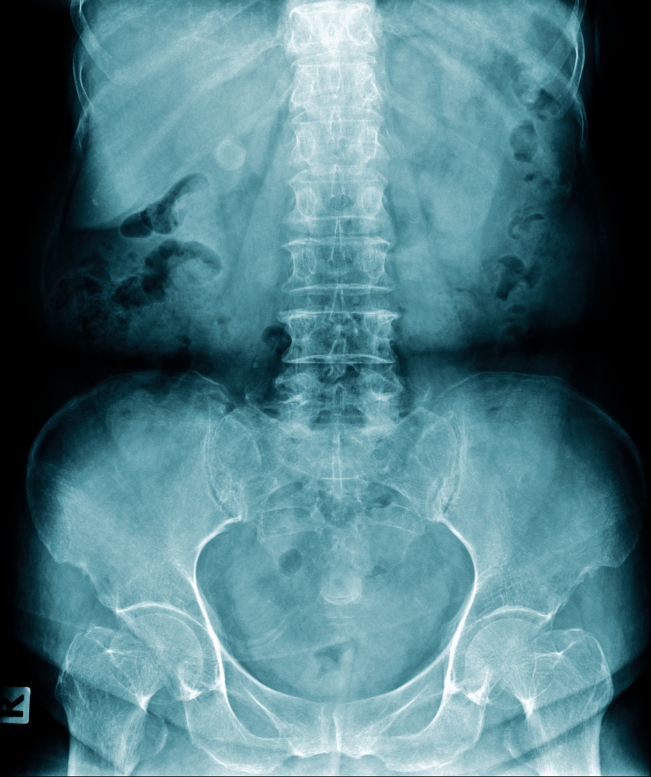
Generally, only an X-ray of the lumbar spine is needed to determine a diagnosis of spondylolysis. A SPECT (Single-Photon Emission Computerized Tomography) scan can identify an acute stress reaction in the pars interarticularis. A negative x-ray and positive SPECT bone scintigraphy would suggest further imaging is needed. A CT scan or MRI can get a better image of the back, while detecting small fractures or ruling out other spine conditions.