Pain
Diagnosing Thoracic Outlet Syndrome (TOS)

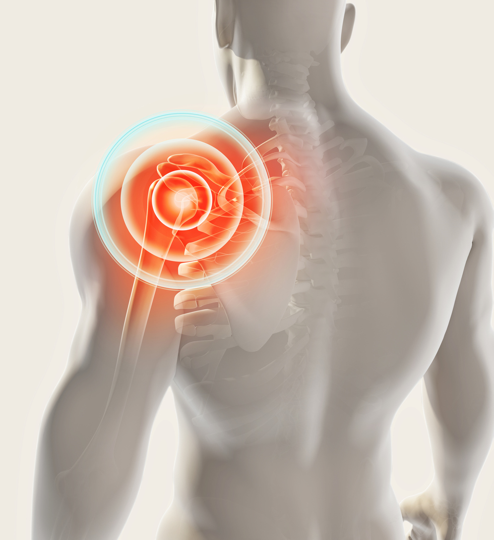
What is thoracic outlet syndrome (TOS)?
Thoracic outlet syndrome (TOS) is a condition in which the nerves and/or blood vessels in the thoracic outlet become compressed, irritated or injured. The thoracic outlet is a narrow space located between the collarbone and the first rib. The nerves and blood vessels that move down the arm, and the muscles from the neck to the shoulder, run through the thoracic outlet.
Diagnosing thoracic outlet syndrome
Symptoms and severity of TOS vary greatly, causing difficulty in obtaining a diagnosis. A health care professional will collect a medical history, perform a physical examination, and complete additional tests as needed. A neurologist may also complete a thorough examination to rule out cervical spine disease or other neurological conditions. A physician may give an injection to relax the muscle to see if symptoms disappear.
Medical history
The physician will ask about medical history, symptoms, occupation, and physical activities. Symptom information will be discussed in depth, such as type of pain, when it began, if anything helps or hurts, severity, etc.
Physical examination
A health care professional will check for physical signs, such as abnormality of the collarbone, swelling or discoloration in the arm, irregular pulse, a depression in the shoulder, etc. Typically, range of motion will be tested by moving and lifting the arm and turning the head. The Adson’s maneuver involves moving the shoulder joint to recreate pinching of the nerve.
Imaging
A health care professional may order certain imaging tests to confirm a diagnosis of TOS, which may include the following:
- An Ultrasound may be the first imaging test performed to assist in diagnosing TSO. It includes sound waves that create images of the body to check for vascular thoracic outlet syndrome or other vascular problems.
- An X-ray of the chest may be ordered to check for an extra rib (cervical rib). It can also help rule out other conditions.
- A computerized tomography (CT) scan includes an injection of a dye into a vein to view blood vessels. It uses X-rays to obtain cross-sectional images of the body. A CT scan can identify the cause and location of blood vessels that are compressed.
- Magnetic resonance imaging (MRI) uses radio waves and magnets to create a detailed view of the body to determine the cause and location of blood vessels that may be compressed. Sometimes, a dye is injected to see a more detailed view of the blood vessels. An MRI may also reveal a fibrous band connecting the spine to the rib or a cervical rib. The head, shoulders and neck may be placed in various positions for a better view of the blood vessels in the arm.
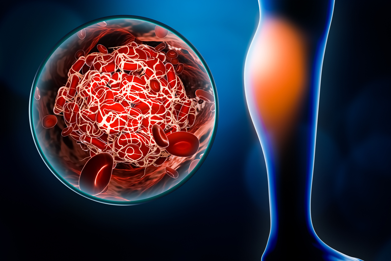
- An arteriography and venography consists of the insertion of a thin, flexible tube through a small incision, normally in the groin. The tube (catheter) is moved through major arteries or through the veins to the affected blood vessels. A dye is injected through the tube to show X-ray images of the arteries or veins. If a vein or artery has a clot, medication can be administered through the tube to dissolve the clot.
- An electromyography (EMG) involves a health care professional inserting a needle electrode into various muscles to check for electrical activity when muscles contract and when they are at rest. This can determine if nerve damage is present.