Pain
Diagnosing Traumatic Brain Injury

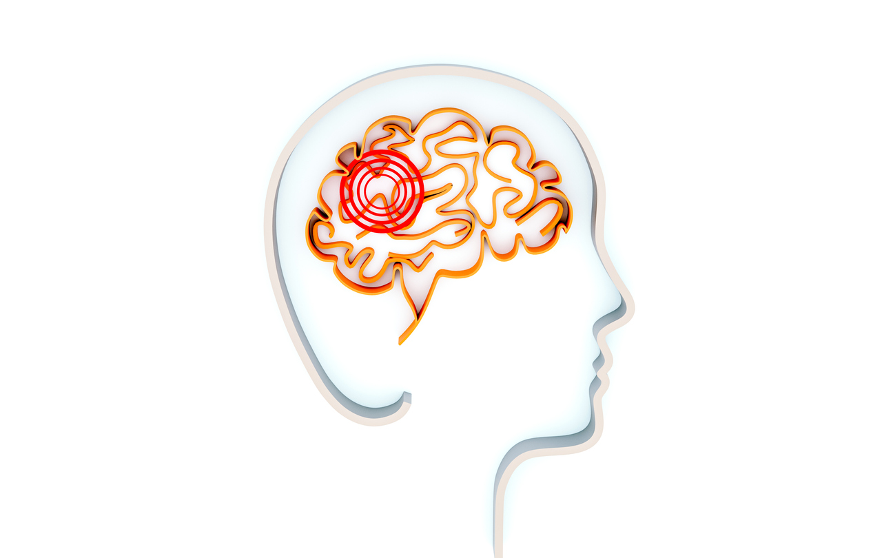
What is a traumatic brain injury?
A traumatic brain injury (TBI) is a type of brain damage that occurs as a result of an injury to the head. This type of injury may be non-penetrative, such as a blow to the head, or penetrative, such as a gunshot wound. The severity of a TBI depends on various factors, and the lasting effects can range from a few days to permanent brain damage or, in severe cases, death. A concussion is the most common type of traumatic brain injury.
Diagnosing a traumatic brain injury
Immediate medical assistance is needed when a TBI occurs. Consequences of a severe TBI can worsen rapidly. A proper diagnosis typically includes a medical history, physical examination, Glasgow Coma Scale, imaging tests, and intracranial pressure monitoring.
Medical history and physical examination
The first step in diagnosing a TBI is a physical exam. The medical professional will assess the physical state and ask questions regarding the injury. Information needed may include the following:
- Was consciousness lost? If so, for how long?
- How did the injury occur?
- What is the location where the head or body was struck?
- Was the body jerked around?
- What was the force of the injury?
- What changes have been observed in speech, coordination, etc.
Glasgow Coma Scale
The Glasgow Coma Scale is a useful tool in assessing and diagnosing a traumatic brain injury. This assessment determines how well directions can be followed, movement of eyes and limbs, and coherent speech. The scale is scored from three to 15, with 15 indicating less severe injuries.
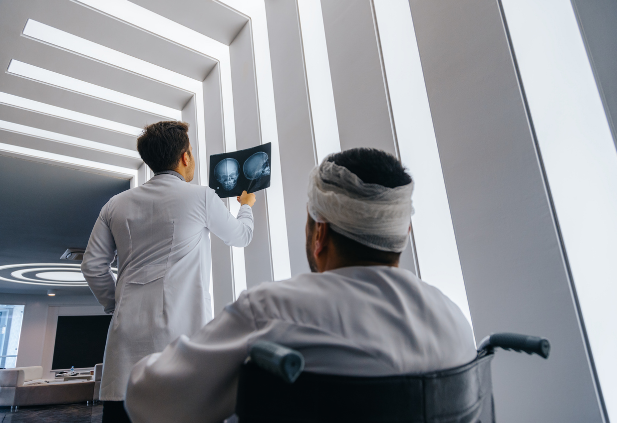
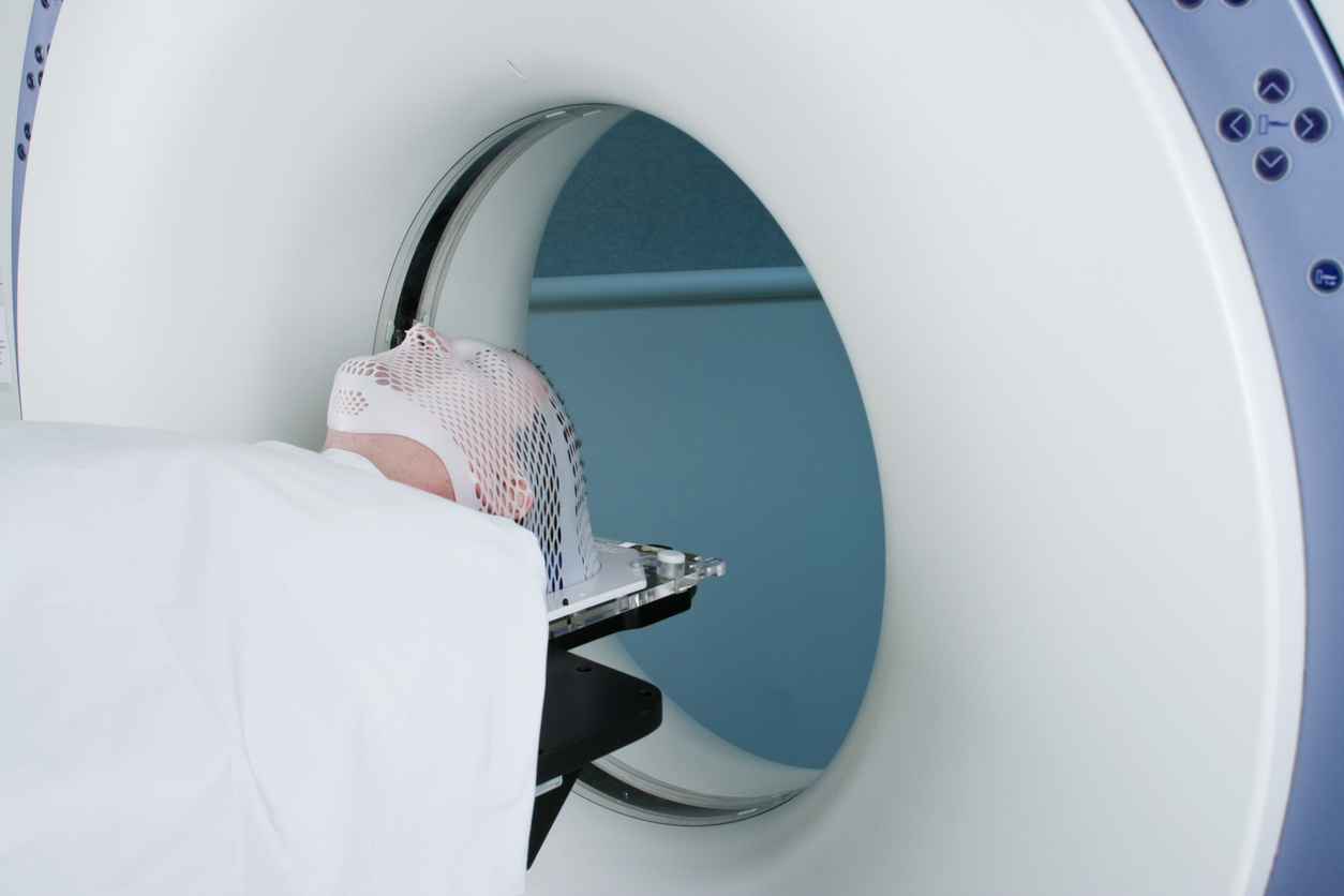
Imaging tests
Certain imaging tests of the brain will likely be ordered. They may include a CT scan or MRI of the brain.
- CT Scan
A computerized tomography (CT) scan utilizes several X-rays to make a detailed map of the brain. It can detect fractures, hemorrhages, hematomas, contusions and swelling in the brain. This is typically the first test that is performed when a TBI is suspected. - MRI
A magnetic resonance imaging (MRI) is a scan that uses magnets and radio waves to create an image of the brain. An MRI is usually performed after the TBI is deemed stable or if brain function does not improve soon after the injury.
Intracranial Pressure Monitor
If a TBI causes tissue swelling, it may result in an increase of pressure inside the skull. This pressure can further damage the brain after the initial injury. In order to monitor the pressure inside the skull, a health care professional may use an intracranial pressure monitor (probe) through the skull. The probe then sends information to a recording device.