Pain
Diagnosing Peripheral Vascular Disease (PVD)

What is peripheral vascular disease?
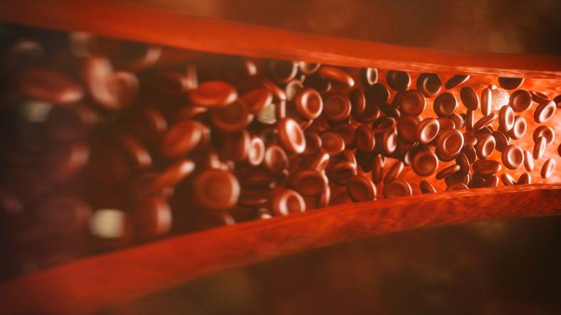
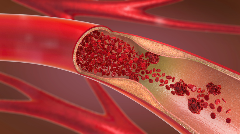
Peripheral vascular disease (PVD) is a progressive circulatory condition that involves the narrowing, blockage, or spasms of blood vessels located outside the heart. It is sometimes referred to as peripheral artery disease (PAD); however, peripheral artery disease is a specific type of peripheral vascular disease that only affects the arteries. Peripheral vascular disease is an umbrella term for circulatory conditions that affect the arteries, veins, or lymphatic vessels. Because PVD involves the narrowing, blockage, or spasms of blood vessels, blood flow is restricted in the affected vessel which reduces the transport of oxygen and nutrients to specific areas of the body. The affected area of the body depends on the location of the narrowed or blocked vessel(s); however, PVD most commonly affects the limbs, especially the legs. The most common symptom is pain and cramping in the affected limb.
Diagnostic tests
The diagnostic process generally begins with a physical exam and a personal/family medical history. The physical exam includes checking for a weak pulse in the extremities and changes in nail and skin color due to tissue ischemia. Because 50% of individuals with PVD are asymptomatic, various medical tests may be used during the diagnostic process.
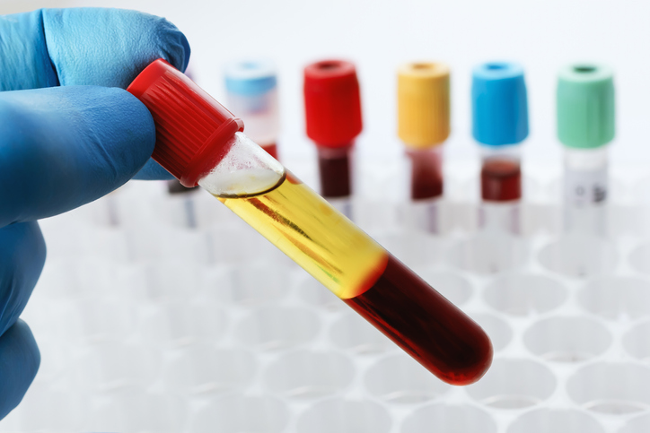
- Blood lipid profile
A blood lipid profile is a blood test that measures levels of fat in the blood (total cholesterol, LDL and HDL cholesterol, and triglycerides). High cholesterol is a contributing factor of PVD. - Ankle-brachial index
An ankle-brachial index (ABI) involves the use of a Doppler ultrasound device and a blood pressure cuff to measure blood pressure in the ankle and in the arm. The two systolic (top) numbers are then compared. The ABI is determined by dividing the blood pressure in the ankle by the blood pressure in the arm. Individuals with severe PVD in the legs present with lower blood pressure in the ankles than in the arms. An ABI of ankle-brachial index of 0.9 to 1.3 is considered normal, less than 0.9 suggests presence of PVD in the legs, and less than 0.5 typically suggests severe PVD in the legs. - Photoplethysmography
A photoplethysmography (PPG) test is similar to an ABI. A small blood pressure cuff is placed around a toe to measure blood pressure, and a PPG sensor (an infrared device) is used to evaluate blood flow near the skin’s surface. Like the ankle-brachial index, the measurement is compared with the systolic blood pressure measurement in the arm. - Doppler ultrasound flow studies
Doppler ultrasound flow studies are non-invasive tests that use high-frequency sound waves and a computer to create images of blood vessels, tissues, and organs. An ultrasound device is used to measure and assess blood flow. Images are created to detect any abnormal blood flow within vessels in the upper thigh, knee, ankle, and arm to determine if any narrowing or blockages of blood vessels are present. - Stress test
A stress test involves the use of a treadmill to elevate the heart rate while a health care provider monitors blood circulation with an electrocardiogram (EKG). If walking on a treadmill is not possible, medication can be used to elevate the heart rate. - Reactive hyperemia test
A reactive hyperemia test can also be used for those who cannot walk on a treadmill. Blood pressure measurements are taken on the thighs and ankles to compare the difference in measurements between the two. - Magnetic resonance angiography
Magnetic resonance angiography (MRA) is a type of magnetic resonance imaging (MRI) procedure. An MRA involves the injection of contrast dye so that blood vessels are more visible during an MRI scan. - Angiography
An angiography is used to detect any narrowing or blockages of the blood vessels in the limbs. It is an X-ray procedure that involves the use of contrast dye injected into an artery. This test is considered the most accurate diagnostic test for PVD as it detects the location, severity and collateral circulation of the arteries. Because of potential adverse side effects, X-ray angiography is used only for severe cases of peripheral vascular disease that are being considered for surgery or angioplasty.