Pain
Diagnosing Sarcoidosis

What is sarcoidosis?
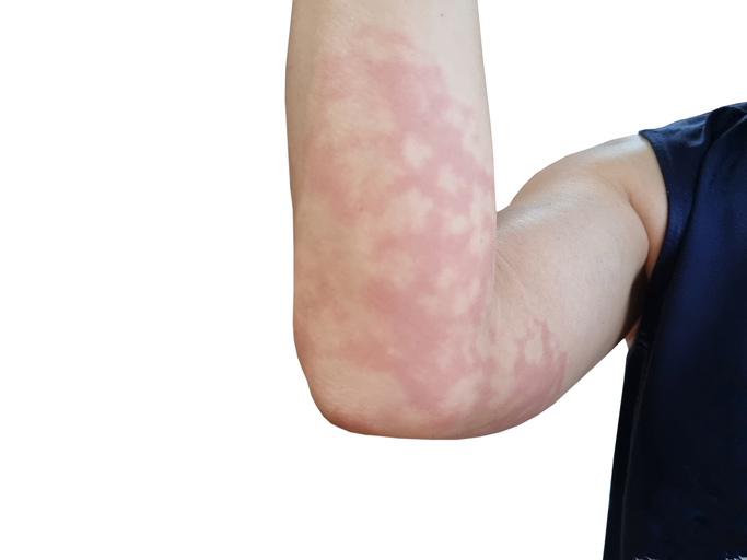
Sarcoidosis is an inflammatory disease characterized by the formation of small collections of inflammatory cells (granulomas) in one or more organs. These growths most commonly form in the lungs and lymph nodes, but they can also develop in the skin, eyes, musculoskeletal system, nervous system, heart, liver, or kidneys.
How is sarcoidosis diagnosed?
Diagnosing sarcoidosis can be a challenge. Because symptoms are not usually present during the early stages of the condition, and some symptoms of sarcoidosis overlap with symptoms of other health conditions, the diagnostic process can be complicated. Sarcoidosis is also categorized into several types. Depending on the type, different organs and systems of the body are affected, which means sarcoidosis presents with a wide range of symptoms that differ from person to person.
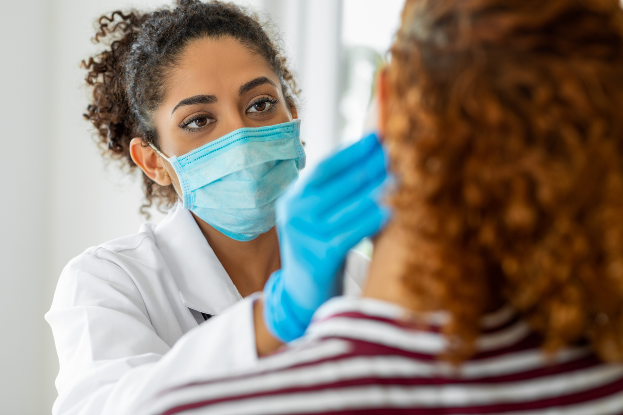
The diagnostic process typically begins with a medical history and physical exam. The physical examination involves looking for common symptoms of sarcoidosis including skin bumps, swollen lymph nodes, and an enlarged liver or spleen. The health care provider also listens to the heart and lungs. Depending on the information collected from the physical exam and medical history, a variety of other medical tests may be ordered.
Chest X-rays
X-rays of the chest are used to look at the heart, lungs, and surrounding tissues, checking for swollen lymph nodes, scarring, or granulomas.
CT scan
CT scans lend a more detailed look at the chest and lungs than chest X-rays. They can also provide a more in-depth look at granulomas and swollen lymph glands.
Pulmonary function (breathing) tests
Pulmonary function tests can determine how well the lungs are transporting oxygen to the blood, how well the lungs are expanding, and how much air the lungs can move. In cases of sarcoidosis, granulomas and fibrosis reduce lung capacity and disrupt the normal exchange of oxygen and carbon dioxide between the lungs and the blood.
Bronchoscopy
A bronchoscopy involves the insertion of a small tube down the trachea and into the bronchial tubes of the lungs in order to inspect the bronchial tubes, rule out infection, and to do a bronchoalveolar lavage.
- Endobronchial ultrasound (EBUS) may be done during a bronchoscopy. The ultrasound probe guides a needle inserted through the bronchoscope in order to biopsy a lymph node. This test can provide a definitive diagnosis of sarcoidosis if it is present.
- Bronchoalveolar lavage involves the use of a bronchoscope to enter the lungs and “wash out” cells and other material from the air sacs. The fluid is then tested for the amount of various cells and other substances that suggest inflammation and immune activity in the lungs.
Gallium scanning
A gallium scanning is a nuclear medicine test; it involves the injection of the radioactive chemical element gallium-67 into a vein. The gallium collects in parts of the body that are affected by immune conditions. Two days after the injection of gallium-67, the body is scanned, and areas with an increased uptake of gallium indicate that inflammation is present. This test does not provide a definitive diagnosis of sarcoidosis; it simply indicates inflammatory activity. Because of this limitation, it is rarely used today.
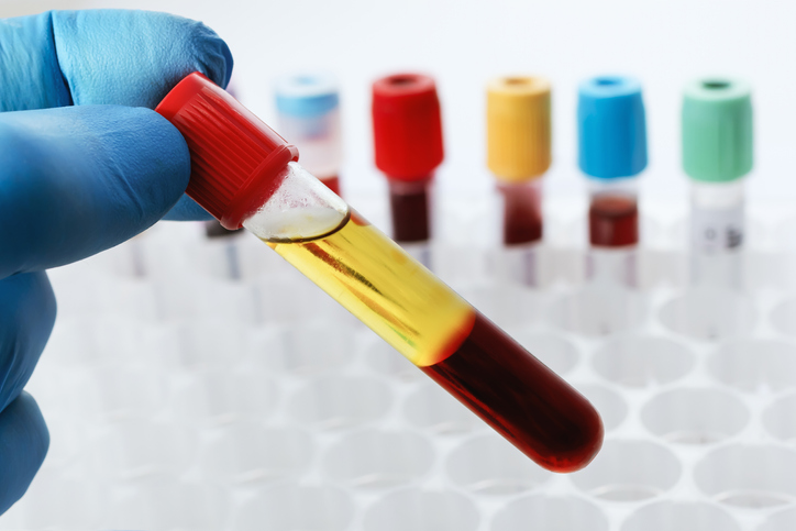
Blood tests
Blood tests analyze the body’s overall functioning, evaluate the number and types of blood cells and blood proteins in the body, as well as examine the health of the liver. One specific blood test measures the level of angiotensin converting enzyme (ACE); granulomas secrete ACE, so high levels of ACE may indicate the presence of sarcoidosis. However, this blood test is not a definitive test as some individuals with sarcoidosis do not show high levels of ACE, and ACE levels can be elevated with other health conditions. A serum calcium test may also be done as increased serum calcium levels often accompany sarcoidosis. A newer blood test that is often done instead of the ACE test is soluble interleukin 2 receptor levels (sIL2R).
Urine test
A urine test can be used to check kidney function and any excess calcium in the urine.
Electrocardiogram (ECG)
An ECG can detect heart abnormalities, such as arrhythmias and palpitations, that may be caused by the presence of scarring or granulomas.
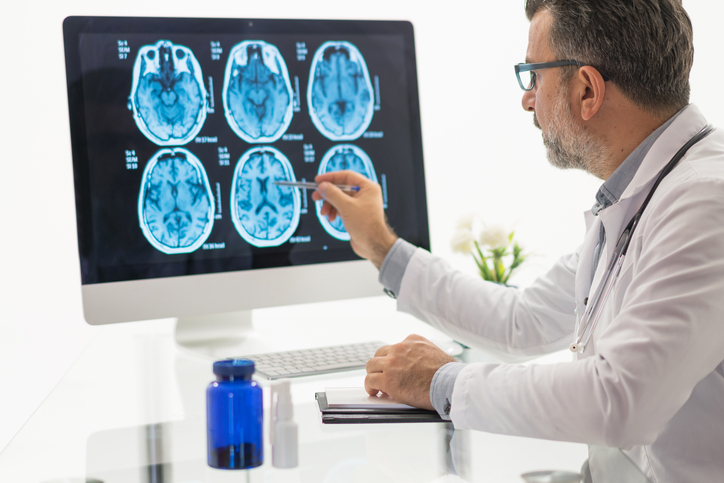
Magnetic resonance imaging (MRI) scan
An MRI may be used to check if sarcoidosis is affecting the heart or nervous system.
Positron emission tomography (PET) scan
A PET scan involves the use of radioactive glucose to identify possible granulomas. While granulomas absorb the glucose, inflammation associated with other health conditions also absorb the material, so a PET scan is often used in conjunction with other tests for sarcoidosis.
Slit lamp examination
A slit lamp examination is a specific type of eye exam that allows a health care professional to look inside the eyes to check for sarcoidosis-related eye damage.
Kveim test
A Kveim test involves the injection of a standard preparation of sarcoid tissue material into the skin. If a lump forms at the injection site, it is considered positive for sarcoidosis, but false positives happen. This test is not currently used in the United States as the FDA has not approved any test material. It is possible for hospitals or labs to have the material prepared for their own use.
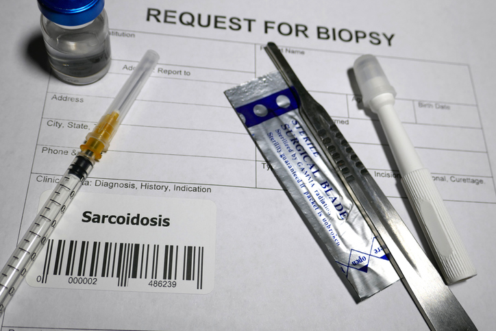
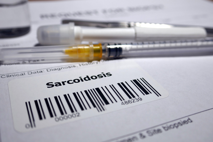
Biopsy
A biopsy involves the removal and testing of a small amount of tissue. Depending on the suspected affected area, biopsies are taken from the skin, lungs, or lymph nodes.