Pain
Diagnosing Scleroderma
What is scleroderma?
Scleroderma is an autoimmune condition that involves hardening and tightening of the skin and connective tissues. There are two main types of scleroderma: localized and systemic. Localized scleroderma usually affects the skin only. Systemic scleroderma affects the skin, underlying connective tissues, or major organs.
Diagnostic process
A diagnosis of scleroderma is made by a clinician based on evidence from a combination of a physical exam and various studies, such as lab tests and imaging tests.
Physical examination
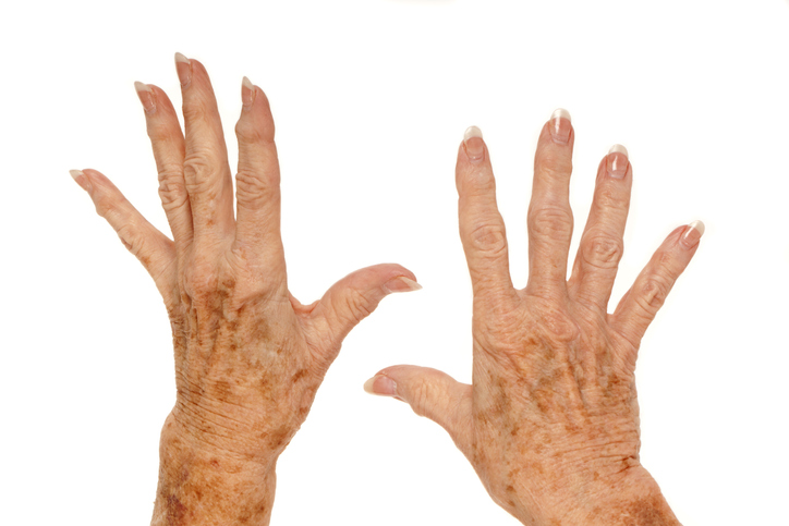
A physical exam can reveal signs that indicate a diagnosis of scleroderma. Some of these clinical clues include the following:
- Evidence of skin thickening may be present on the hands, arms, toes, legs, or face. This may include a condition called sclerodactyly (thickening of skin on the hands).
- Ulcers on fingers and toes can indicate poor circulation. This can occur with Raynaud’s phenomenon, which is associated with limited scleroderma. Raynaud's is a disease that decreases blood flow to extremities in response to cold.
- Calcium deposits under the skin is also a frequent finding of scleroderma.
Laboratory tests
Laboratory tests include blood tests, urine tests, and skin biopsies.
- Blood tests are used to check for elevated immune factors, called antinuclear antibodies, which are found in over 90% of scleroderma cases.
- Blood tests and urine tests are performed to check the function of the kidneys.
- Skin biopsies may reveal calcium deposits under the skin, changes to skin blood vessels, or excessive production of collagen (all signs of scleroderma). One major limitation of skin biopsies is that they cannot distinguish between localized and systemic scleroderma.
Pulmonary function tests (PFTs)
Pulmonary function tests examine how well the lungs are functioning. Systemic scleroderma often presents with lung involvement which may include lung scarring or pulmonary hypertension.
Imaging studies
Imaging studies, such as x-rays, CT scans, and MRIs, can be used to show and monitor changes in the bones and other tissues that may be caused by scleroderma. A CT scan of the chest can also be used to measure the extent of lung involvement.
Cardiac studies
Electrocardiograms and echocardiograms measure how well the heart is functioning. Scleroderma often causes heart scarring.
- An electrocardiogram, or ECG, is a test that measures the electrical activity of the heart.
- An echocardiogram checks the structure of the heart and can also be used to determine if pulmonary hypertension is present. Pulmonary hypertension is a common complication of scleroderma. In some cases, a holter monitor test may be done, which involves wearing a type of electrocardiogram for 24 to 48 hours.
Gastrointestinal studies
Gastrointestinal studies are performed to check for evidence of scleroderma as it can affect the muscles of the esophagus and intestines, leading to heartburn/acid reflux, swallowing difficulties, and problems with the absorption and movement of food in the intestines. Possible tests include the following:
- Endoscopy to view the digestive tract
- Manometry test to see if the esophagus is working properly
- Barium study to diagnose swallowing problems
- Gastric emptying study to test how food moves out of the stomach
- Esophageal pH monitoring to evaluate for possible acid reflux