Pain
Diagnosing Spondylolisthesis

4 people found this helpful
Print
Share
Save
What is spondylolisthesis?
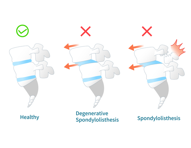
Spondylolisthesis is a medical condition that primarily affects the lower back (lumbar spine); however, it can also occur in the mid to upper back (thoracic spine) or neck (cervical spine). The body has 33 stacked vertebrae that form the foundation of the spine. Spondylolisthesis occurs when a vertebra slips forward (anterolisthesis) or backward (retrolisthesis) onto the vertebra underneath it, which may lead to nerve compression.
Diagnosing spondylolisthesis
A health care professional will obtain a medical history, perform a physical examination, and complete necessary imagining to diagnose spondylolisthesis.
- Medical History. A physician will ask about symptoms to get a clear idea of what an individual is experiencing. This will include when pain began and its intensity.
- Physical Examination. A physical examination will be performed to test for symptoms of spondylolisthesis. This may involve stretching the leg straight outward, which is difficult with spondylolisthesis.
- Imaging. Certain imaging tests can confirm a spondylolisthesis diagnosis. An x-ray of the spine can determine if a vertebra has moved. A CT scan or MRI provides a detailed view of the spine to observe soft tissue, such as the discs and nerves. They also determine if a displaced vertebra is impinging on a nerve.