Pain
Diagnosing Back Pain

390 people found this helpful
Print
Share
Save
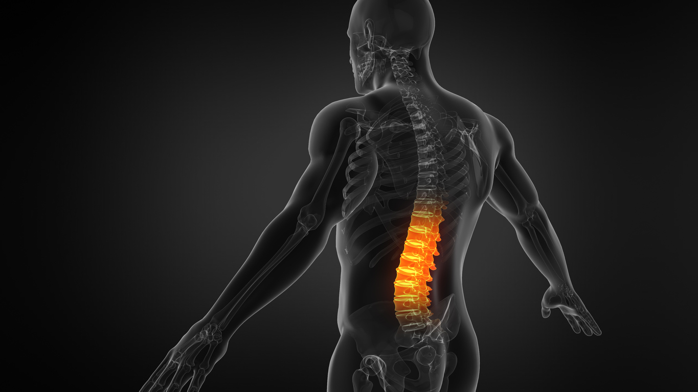
Back pain is the most common reason for medical center visits. An estimated eight out of ten people will experience back pain at some point. It can appear suddenly or develop gradually. Symptoms range from mild to severe.
How is back pain diagnosed?
Back pain is caused by various conditions. In order to receive appropriate treatment, a health care professional will need to make a correct diagnosis. This includes gathering a medical history, performing a physical examination, and ordering tests.
- Medical history. A health care professional will ask about family history. Other questions may include when the pain began, if it was sudden or gradual, does it radiate to other areas, and if heavy lifting was involved. A description of the pain is needed, such as sharp, dull, achy, etc. They will ask about any recent illnesses or additional symptoms.
- Physical examination. A physical exam may include looking for nerve damage during walking, sitting or standing. The reflexes at the knees and behind the ankles are measured. Diagnosis will likely include movement of the hip, knee, big toe and ankle. Leg strength, ability to detect sensation, and range of motion may be evaluated. A thorough palpation along the back can locate muscle spasms, tightness, tenderness, or abnormalities.
- Blood or urine test. Depending on the cause of pain, blood tests or urine tests may be ordered. Blood tests can detect genetic markers, infections, or other conditions. A urine test can check for kidney stones or other underlying illnesses.
- X-ray. An X-ray uses radiation to create images of the bones. It can detect arthritis, broken bones, bone spurs, tumors, or alignment. However, it will not expose issues with discs, nerves, muscles, or the spinal cord.
- CT scan. A computer tomography, or CT scan, uses X-rays and computer systems to create a 3D image to see bones and soft tissues. It assesses blood vessels, muscles, nerves, ligaments, and discs. A myelogram is a CT scan that includes a dye injection around the nerve roots to highlight spinal structures.
- MRI. A magnetic resonance imaging scan, or MRI, uses a magnet and radio waves to provide a detailed image of spinal structures. It can detect abnormalities with the muscles, ligaments and discs. An MRI can also locate misalignment or joint overgrowth.
- Electromyogram. An EMG involves inserting tiny needles into the muscles to monitor electrical activity. It checks nerves and muscles for nerve damage. This test can also confirm nerve damage due to herniated discs or spinal stenosis.
- Bone scan. Although rare, a bone scan may be ordered to look for compression fractures or bone tumors. It involves using a small amount of radioactive substances, a computer, and a special camera to look closely at the bones.