Pain
Diagnosing Lymphedema
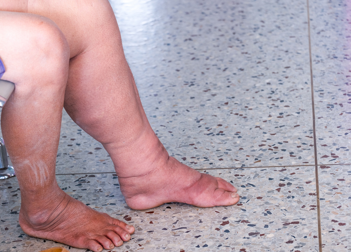
What is lymphedema?
The lymphatic system moves a colorless, watery fluid (lymph) throughout the body to help filter waste and fight off pathogens. Lymphedema develops when the lymphatic system is unable to adequately drain this fluid. Because it is not being properly drained, the fluid accumulates in the surrounding soft tissues, causing swelling and various other symptoms. Lymphedema can occur anywhere in the body but most commonly occurs in the arms and legs.
Diagnosing lymphedema
The diagnosis of lymphedema is determined based on findings from a physical examination, medical history, and medical tests.
Physical exam
A physical examination can reveal signs that indicate a potential diagnosis of lymphedema. Some of these signs include the following:
- Pitting edema or a cobblestone appearance of the skin (papillomatosis)
- Lymph fluid leaking from the skin (lymphorrhea)
- The inability to pinch a fold of skin at the base of the second toe (Stemmer’s sign)
Medical history
A medical history can provide clinical clues of lymphedema. Along with findings from a physical exam, a diagnosis of lymphedema may be suspected based on the following:
- A family history of lymphedema
- A causative event, such as a surgery, cancer treatment, or trauma
- Comorbidities such as Noonan syndrome, Turner syndrome, or a high body mass index (BMI)
Medical tests
Medical tests that may be ordered during the diagnostic process include the following:
- Bioimpedance spectroscopy
A bioimpedance spectroscopy involves sending a weak electrical current through the body to measure the amount of fluid present in tissues. This test can help determine if lymphedema is present. - Lymphoscintigraphy
A lymphoscintigraphy involves radionuclide imaging of the lymphatic system. The body is scanned after radioactive dye is injected. The images show the dye moving through the lymphatic system and highlight any blockages. - Doppler ultrasound
A Doppler ultrasound monitors blood flow and pressure, helping to identify any blockages. This test can also help rule out blood clots and venous abnormalities that may be mistaken for lymphedema. - Computerized axial tomography (CT) scan
A CT scan uses X-ray technology to create computer-generated images of bones, organs, and other tissues. CT scans can be used to find obstructions and eliminate other potential causes of lymphedema. - Lympho-MRI (magnetic resonance imaging)
A lympho-MRI is a medical imaging test that uses a magnetic field and computer-generated radio waves to provide detailed images of the entire lymphatic system. - Lymphatic mapping with fluorescence navigation
Lymphatic mapping with fluorescence navigation involves the injection of a fluorescent dye into the web of a hand or foot to visualize the superficial lymphatic system in real time.