Pain
Diagnosing Temporomandibular Joint (TMJ) Disorder
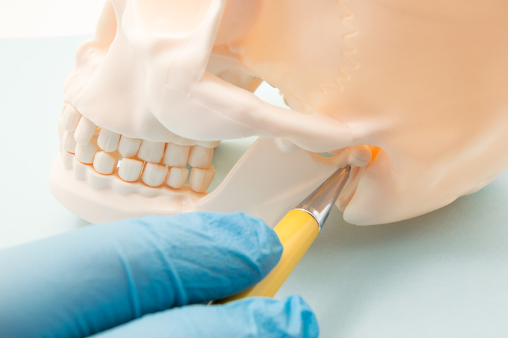
What is temporomandibular joint disorder?
The temporomandibular joint (TMJ) joins the lower jaw to the skull; it is located in front of each ear on both sides of the head. It acts as a hinge and allows the jaw to open and close and move from side to side. The bones in the joint are covered with cartilage and are separated by a small disc, which allows for smooth movement of the jaw.
Temporomandibular joint disorder is characterized by pain in the jaw as well as the nerves and muscles surrounding the jaw. Temporomandibular joint disorder is often referred to as simply “TMJ” in reference to the affected joint.
Diagnostic process
If not properly treated, TMJ disorder can worsen and potentially lead to permanent injury. A visit with a primary care physician, dentist, oral surgeon, or TMJ specialist for a proper diagnosis and treatment plan is important.
TMJ disorder is often difficult to diagnose because various other health conditions, such as tooth decay, ear infections, facial nerve pain, sinus issues, gum disease, tumors, and arthritis pain, can cause similar symptoms. The diagnostic process includes a medical history, physical exam, and imaging tests.
- Medical history
A medical history for TMJ typically includes questions about any recent injuries, other health conditions, and current medications. Questions about any family history of TMJ may also be asked. - Physical exam
A physical exam for TMJ consists of a health care professional examining the joints of the jaw for areas of pain, tenderness, or swelling; listening for any popping, grating, or clicking noises that the jaw produces as it opens and closes; and observing the range of motion of the jaw as it is moved. The face, ears, mouth, throat and neck are also examined. - Medical imaging
X-rays are commonly ordered to view the jaw bones, temporomandibular joints, and teeth. Other imaging tests, such as a CT scan or MRI, may also be ordered. A CT scan can provide more detailed images of the temporomandibular joint, and an MRI can reveal if the disc in the temporomandibular joint is in the proper position as the jaw moves.