Pain
Diagnosing Primary Lateral Sclerosis (PLS)

What is primary lateral sclerosis (PLS)?
Primary lateral sclerosis (PLS) is a rare neuromuscular disorder that involves the gradual degeneration of nerve cells —more specifically, upper motor neurons — in the brain. Upper motor neurons are responsible for the initiation of voluntary movements and help with body posture. PLS belongs to a group of conditions known as motor neuron diseases.
How is PLS diagnosed?
No specific test exists that can confirm a diagnosis of PLS. The diagnosis of PLS is usually aided by an individual’s medical history, clinical presentation, and medical tests to rule out other medical conditions. PLS may be diagnosed based on the presence of the following:
- Age ≥ 25 years
- Progressive upper motor neuron dysfunction ≥ 2 years
- Signs of upper motor neuron dysfunction in two of three regions: lower extremity, upper extremity, or brain stem
Along with the absence of the following:
- Sensory symptoms (unexplained by a comorbid condition, such as diabetes)
- Active lower motor neuron disease
- An alternative diagnosis that explains upper motor neuron dysfunction
Medical tests
PLS can mimic signs and symptoms of other diseases such as multiple sclerosis and amyotrophic lateral sclerosis (ALS). Tests that may be ordered to rule out other medical conditions include the following:
- Blood work
Blood tests are often used to assess general health and to rule out other health conditions, such as infections, that may be the cause of symptoms. - Electromyography (EMG)
An EMG is a test in which a small needle is inserted into a muscle to measure and evaluate electrical activity in the muscle. This test can be used to determine if there is lower motor neuron involvement. Lower motor neurons begin in the spinal cord and innervate the muscles and glands in the rest of the body. This test can be used to differentiate primary lateral sclerosis from amyotrophic lateral sclerosis. - Nerve conduction study
A nerve conduction study is a test in which two electrodes are placed on the skin to measure the speed of electrical impulses. It assesses a nerve’s ability to send messages to muscles throughout the body. This test can help rule out other muscle or nerve diseases. - Lumbar puncture (spinal tap)
A lumbar puncture involves the careful insertion of a needle into the spinal canal via the lower back. A small amount of cerebrospinal fluid is removed for testing. This is often done to rule out infections or other inflammatory conditions that may be the cause of muscle weakness. - Magnetic resonance imaging (MRI)
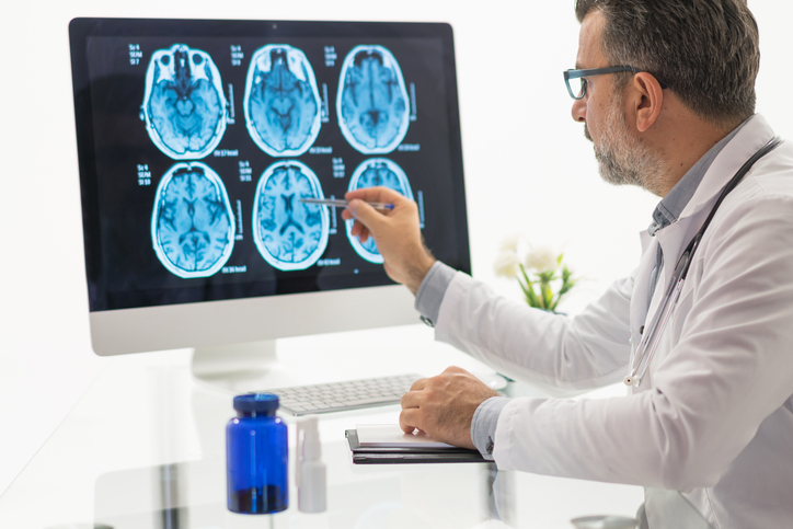
An MRI of the brain or spine may reveal nerve cell damage. It can also help rule out other health conditions, such as tumors or multiple sclerosis, that may be the cause of symptoms.